


cambridge, ma
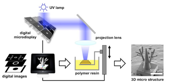
In projection micro-stereolithography, UV light passing through a modified digital projector is focused down to a tiny region. The resulting sub-micron resolution suits this technology well for biological applications. The system we designed and built was used at MIT to create vascularized scaffolds for liver cells growth.
In projection micro-stereolithography, UV light passing through a modified digital projector is focused down to a tiny region. The resulting sub-micron resolution suits this technology well for biological applications. The system we designed and built was used at MIT to create vascularized scaffolds for liver cells growth.